Strictly speaking, these are protists, rather than plants, but
many people still think of them as plants: after all they do possess chlorophyll
for photosynthesis. Of course many have the disconcerting habit of moving
around, often quite rapidly, and not very plant-like...
Such slides were especially popular in the 19th century as exhibition
pieces to show of both the beauty of the diatoms and also the prowess of the
slide maker. It has been said that the ideal tool for arranging diatoms is
a tiger's whisker.
|
Diatoms
|
| Diatoms have long been a favourite subject
for microscopists, both for aesthetic and utilitarian reasons. The patterns
resulting from their cell-wall structure can be very beautiful, especially
if you have a love of patterns and geometry. These patterns can be so
fine that they have also provided microscopists with a readily available
and cheap means of testing the ability of their instuments to resolve
fine detail. The pictures on this page of diatoms are of a mixture of
marine and freshwater species.
Diatoms are unusual in that as single celled algae they
possess cell wals impregnated with silica - effectively the cell walls
are made of glass. The cells are made of two halves, "frustules"
held together by a kind of belt, called a "girdle". To appreciate
the fine patterning, the frustules have to be specially cleaned and
mounted for viewing in a highly refractive mountant. The patterns are
only poorly visible in live diatoms, largely being obscured by chlorophyll
and also by the fact that the refractive index of water is too low for
really good resolution.

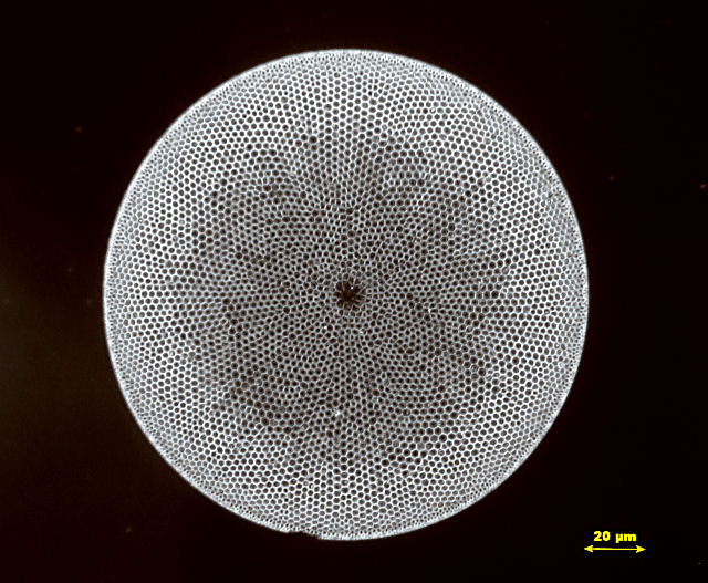
Moiré pattern formed by fine markings in a centric
diatom
Zeiss GFL Microscope
Objective: Zeiss x40 plan achromat
Ocular: Watson x8 compensating
COL with Zeiss 0.9 NA substage condenser
Camera: Canon Powershot S50
ISO100, F4.9. 1/13 sec
Sample from Dunkirk, USA, kindly provided by Frez

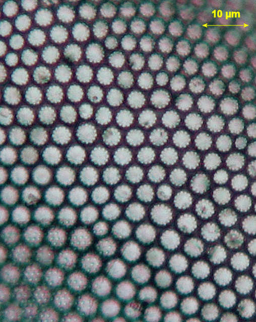
Colouration caused by diffraction effects due to the very fine patterns
Zeiss GFL Microscope
Objective: Zeiss x25 plan achromat
Ocular: Watson x8 compensating
Darkground with Zeiss 0.9 NA substage condenser + wheelstop
Camera: Canon Powershot S50
ISO100, F4.9. 1/8 sec
Sample from Dunkirk, USA, kindly provided by Frez
|
Coscinodiscus
Zeiss GFL microscope
Objective: x25 plan achromat
Ocular: Watson x8 compensating
Zeiss dark field substage condenser
Light source: 8000 mcd LED
Camera: Canon Powershot S50
F4.9, 0.5 sec, ISO50
and at greater magnification: -

Zeiss GFL Microscope
Objective: Zeiss x100 oel
Ocular: Watson x8 compensating
Substage: Zeiss brightfield NA 1.3
Camera: Canon Powershot S50
ISO100, F4.9, 1/160 sec
Another species:
Zeiss GFL Microscope
Objective: Zeiss x40 plan achromat
Ocular: Zeiss x10 Kpl
Brightfield, flash
|
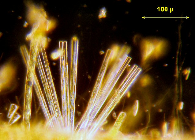
Synedra
A live sample from Dipping Pond 1, Warnham Millpond 25-Apr-2004
Watson Bactil Binocular Microscope
Ocular: x10 Huygenian
Objective: x10 parachromat
DG illumination (wheelstop)
Camera: Canon Powershot S50
|
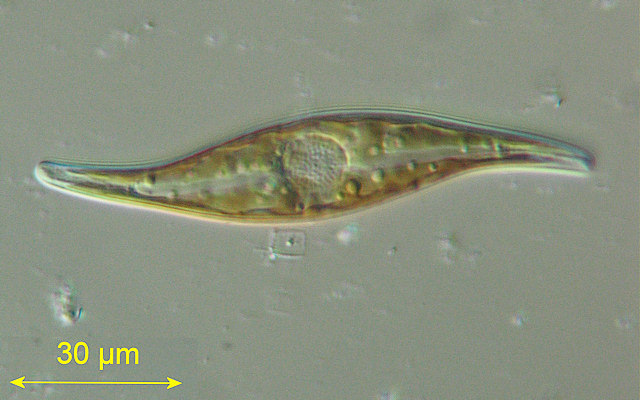
Gyrosigma sp
A common freshwater species
Microscope: Zeiss Standard GFL
Objective: Leitz x40 NPL Fluotar ICT
Ocular: Olympus P15
Substage: Leitz ICT
Sample from Warnham Mill Pond mud

Pleurosigma sp (prepared specimen)
A marine species
Showing detail of fine markings (inset)
Specimen from Dunkirk, USA (thanks, Frez!)
Zeiss GFL Microscope
Objective: Zeiss x40 plan achromat
Ocular: Watson x8 compensating
Circular oblique lighting: Zeis 0.9 NA condenser with substage wheelstop
Camera: Canon Powershot S50
ISO100, F4.9, 1/8 sec
|
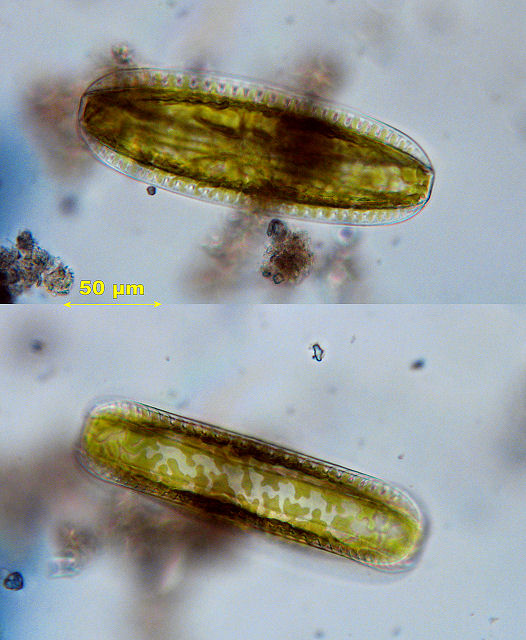
Unidentified diatom
Girdle and plan views (but which is which?)
Zeiss GFL Microscope
Objective: Zeiss x40 Plan achromat
Ocular: Watson x8 compensating
Brightfield
Sample from Warnham Millpond (main body) 03-Jan-2005
|
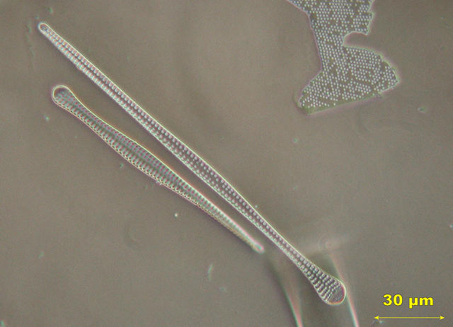
Sceptroneis sp.
Specimen from Dunkirk, USA (also from Frez)
Zeiss GFL Microscope
Objective: Zeiss x40 plan achromat
Ocular: Watson x8 compensating
Circular oblique lighting: Zeis 0.9 NA condenser with substage wheelstop
Camera: Canon Powershot S50
ISO100, F4.9, 1/8 sec
|
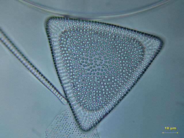
Triceratium sp (?)
Zeiss GFL microscope
Objective: Zeiss x100 oel
Ocular: Watson x8 compensating
Substage: Zeiss NA 1.3 brightfield
Camera: Canon Powershot S50
ISO100, F4.9, 1/160 sec
Combination of 4 images manually
Sample from Dunkirk, Maryland
|
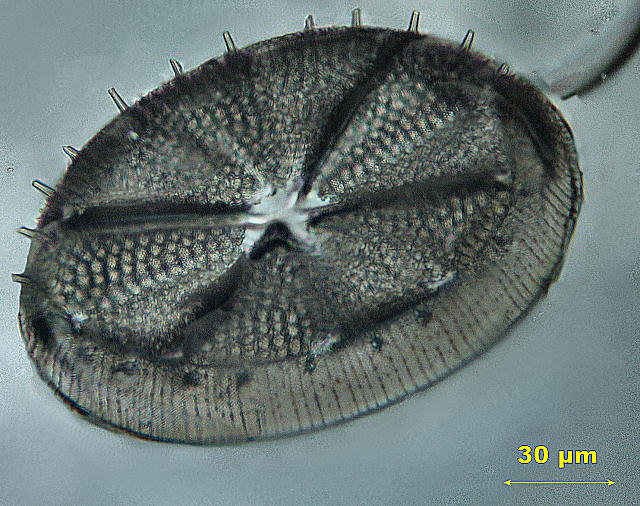
Actinoptychus
This is a composite of 30 images. A rough template was first composed
using CombineZ, then each of the in-focus parts of the original images
was cloned onto the template using Paint Shop Pro 9. The original
specimen is on a slide, provided by Frez, of diatoms from Dunkirk,
Maryland. This particular individual was in the mounting medium at
an angle, thus permitting a composite image showing the 3D layout
of the diatom.
Zeiss GFL Microscope
Objective: Zeiss x40 plan achromat
Ocular: Watson x8 compensating
Substage condenser: Zeiss 0.9 NA brightfield
Camera: Canon Powershot S50
ISO100, F4.9
...and in plan view:

Zeiss GFL Microscope
Objective: Zeiss 40/0.63 plan achromat
Ocular: Zeiss Kpl W 10x/18
Brightfield
Stack of 15 images/Helicon Focus
|
... and finally for Actinoptychus, here is a 3D view again,
but this time created using Helicon Focus to combine 92 differential
interference contrast images:

Microscope: Zeiss Standard GFL
Objective: Leitz 40/0.7 NPL ICT Fluotar
Substage: Leitz ICT
Ocular: Watson x8 Compensating
Stack of 92 images
|

A selection of diatoms in a strew slide of a sample from the Bosphorus
Sea
Zeiss GFL microscope
Objective: Zeiss x40 PH2
Ocular: Watson x8 compensating
Phase contrast
Camera: Canon Powershot S50
ISO200, F4.9, 1/13 sec
|
Desmids
These are another type of single celled algae, and again they come
in many beautiful forms.
|
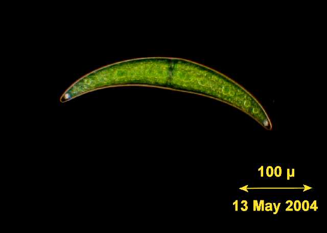
Closterium
Projectina Microscope
Ocular: x10 phot
Objective: x10 achromat
DG illumination: wheelstop
Collected from my garden pond
|
Other Algae

Pandorina
Zeiss GFL Microscope
Ocular: Watson x8 compensating
Objective: Zeiss x40 plan achromat
Brightfield flash
Camera: Canon Powershot S50
ISO100, F4.9, Flash
Sample from Boldings Brook surface film (Warnham Millpond, Hosrham,
UK) 10 July 2005
|

Spyrogyra
Zeiss Standard GFL Microscope
Objective: Zeiss x10 Plan achromat
Ocular: Watson x8 compensating
Brightfield
Sample from Warnham Millpond
1/250 sec, F8, ISO100
Stack of 15 images/Helicon Focus
|
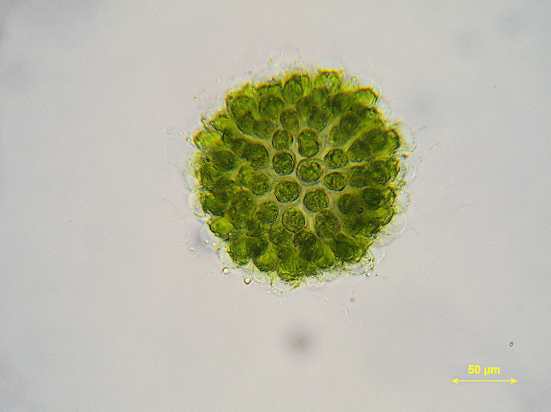
Synura uvella(?)
This is a rather like a spherical cluster of grapes, each one of
which has a flagellum. The whole colony swims with a rolling motion,
tumbling through the water.
Microscope: Zeiss Standard 18
Ocular: Zarf G9 Photo-adapter
Objective: Leica EF L 20/0.3
COL
Camera: Canon Powershot G9/Flash
Sample: Wimbledon Common 31 May 2008
Stack of 12 images, CombineZP
|
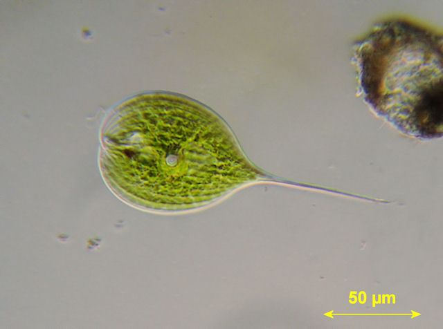
Phacus, a Euglenid alga
Microscope: Zeiss Standard 18
Ocular: Zarf G9 Photo-adapter
Objective: Leica EF L 20/0.3
COL
Camera: Canon Powershot G9/Flash
Sample: Quekett Microscopical Club excursion to Wimbledon Common 31
May 2008
The dark spot at the left end of the organism is in fact a red eye
spot
|

Gonium pectorale
This is a free swimming algal colony, usually of 16 cells, the inset
shows the colony in motion, and the flagella can be seen that provide
propulsion
Microscope: Zeiss Standard GFL
Ocular: Zeiss Kpl W 10x/18
Objective: Leitz x40 NPL Fluotar ICT
Substage: Leitz ICT
Sample from garden pond 11 Aug 2007
|

Coleochaete scutata
Microscope: Zeiss Standard GFL
Ocular: Olympus P15
Objective: Leitz x40 NPL Fluotar ICT
Substage: Leitz ICT
Sample grown in microtube in Pond at Warnham LNR
|
|
|